A Day in Archaeology: developing the NEMCAS (North East Material Culture Analytical Suite) research facility at Durham University
Archaeology blogging feels like almost archaeological at this point! I have run my own archaeology blog, Castles and Coprolites, since 2012. The online world has changed so much in this time. It feels like blogging has been taken over by more short form content and social media, and many people seem to prefer short videos (or ‘vlogging’) over text. But traditional blogging-as-an-online-diary will always be my preferred media. I have taken part in events similar to this in the past (contributing multiple entries to Day of Archaeology which ran 2011 – 2017) but this is my first entry for the CBA Festival of Archaeology. No fieldwork or even lab work for me to talk about today, but I do hope I can give an insight into some of the behind the scenes work that goes into running a research facility that supports all sorts of archaeological research.
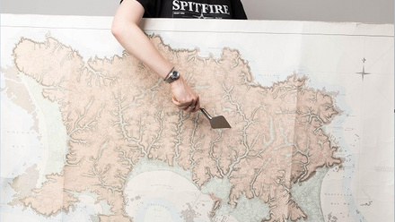
This year, I took a big step in my career, moving to the Department of Archaeology at Durham University after 10 years at Newcastle. My key role will be establishing a new state of the art thin section and XR-CT facility, as part of RICHeS (Research Infrastructure for Conservation and Heritage Science). This is a new 10-year investment from the UK’s AHRC (Arts and Humanities Research Council), to establish a humanities-led distributed research infrastructure across the UK. Thin section micromorphology is a long-established technique in archaeology that I like to describe as ‘excavation under the microscope’. We take intact blocks of soil or sediment from an archaeological site, set them in resin, then cut and grind them into 30 micron slides that can be looked at under the microscope. This lets us observe microscopic features that can tell us how the deposit has formed, including what activities were taking place, and how much disturbance or modifications have happened since the site was in use. It provides a microscale context for all the artefacts and ecofacts contained within the sediment, and is thus an extremely important technique, however there are limitations. Namely that it offers only a 2D snapshot of what are complex 3-dimensional deposits. That is where XR-CT (X-Ray Computed Tomography) comes in. This technique lets us scan a soil/sediment block and provide a 3D image of its internal structure and features.
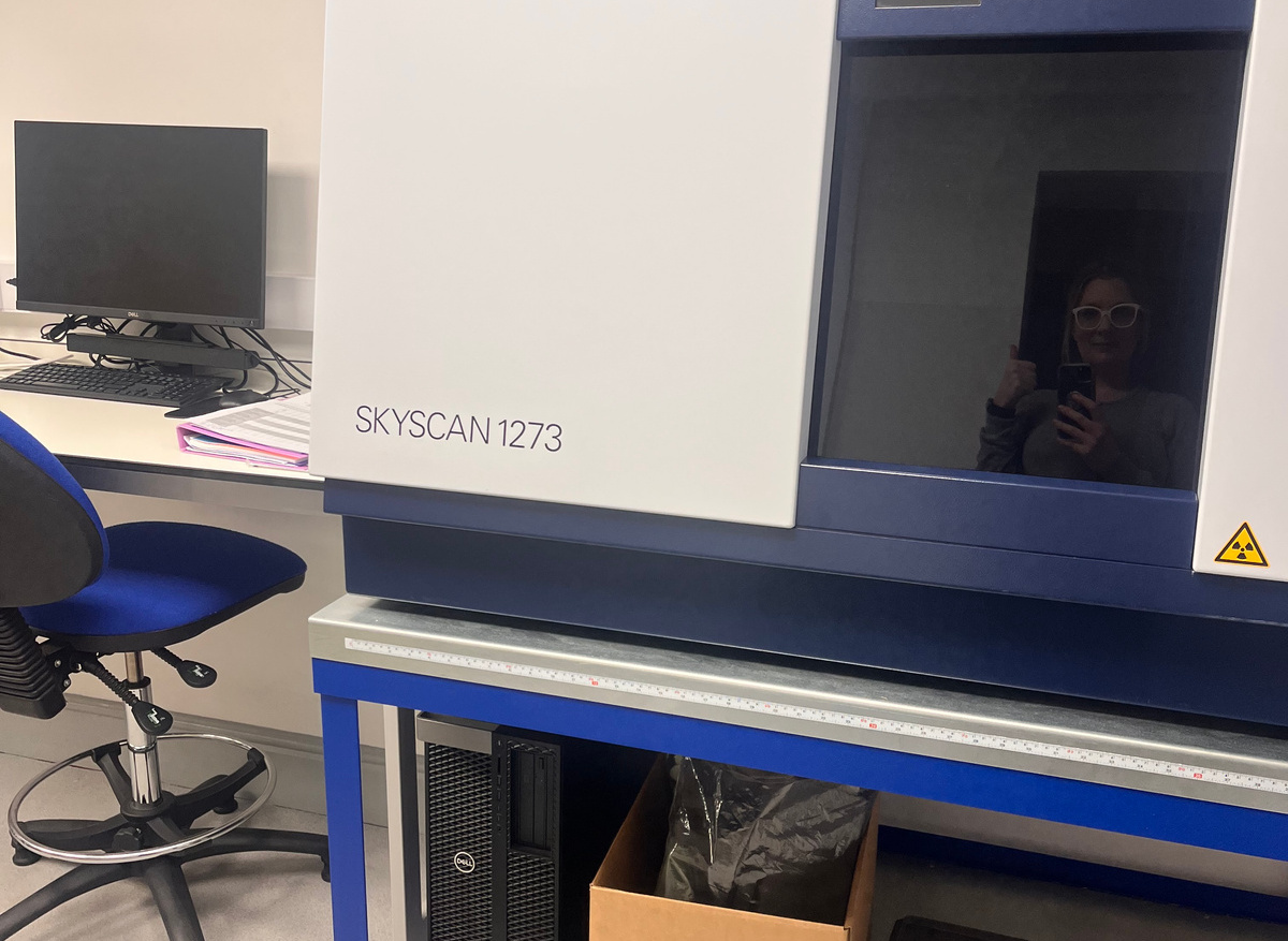
You have probably heard of medical CT scanning, or maybe even had a CT scan yourself – this is a similar technology that scans a whole person to image internal organs and tissues. Our scanner is higher energy than a medical scanner, and quite a bit smaller! We call it a ‘micro-CT’ scanner. It is not exactly small though, it weighs around 450kg and has its own specially made heavy duty bench on castors so it can be moved. Micro-CT works by taking thousands of X-rays of an object from different directions, and constructing a 3D image from this requires high powered computing and complex software. XR-CT has replaced thin section analysis in many areas of soil science, but for archaeology I believe that these two techniques will develop complementary to each other.
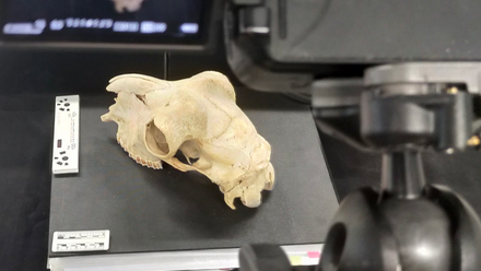
XR-CT is highly versatile, and is also extremely useful for imaging artefacts, letting us have a glimpse inside and to see how these were constructed. We have scanned lots of interesting objects from the Great North Museum, my favourite being an ancient Greek ceramic boar figurine – we were able to ‘look inside’ and could see a fingerprint where the craftsperson pressed the clay into the mould! CT is also really useful for imaging bone, revealing the internal structure of the bone and allowing archaeologists to see features that may relate to stress or disease, for example.
NEMCAS will be the northeast node of RICHeS, and my day-to-day activities at the moment revolve around setting up the facility, promoting it to external researchers, and supporting the development of new projects that will benefit from this amazing equipment. Archaeologists, museum professionals and others will be able to visit the facility and learn how to do these analyses themselves, so setting up training is also on my to do list. Next month I’ll be going to the National Heritage Science Forum Conference to network with other heritage science professionals. Right now I am going to lots of meetings with equipment manufacturers and sales, procurement teams, health and safety, estates teams etc. At the same time I am getting to know my new department and finding out how everything works.
Lisa-Marie Shillito
Professor of Geoarchaeology and Heritage Science